I recently read a 2 decade old editorial on digital imaging in dermatology. The editorial started off with a question:
“Why do dermatologists need any imaging techniques for skin, an entirely visible organ?”

Every time I open my Facebook, I see clinical dermatology images posted for opinion, with a good number of comments and likes. Though crowdsourcing dermatological diagnosis in this manner is a grave violation of privacy to me, digital imaging has become an integral part of dermatology practice.
For some dermatologists (like me), the digital photographs are the only contact with the world of lesions. With the growing popularity of teledermatology and (ethical and non-ethical) crowdsourcing, your exposure to digital photographs as a window to your patient is going to increase. This post is actually a prelude to (an attempt at) defining that future of digital dermatology.
The future has many challenges: Standardization, Sensitivity, Privacy and Ethics are just few of them. In this series, I will discuss these challenges from a technical perspective. I dream of a future when ‘diagnosing from digital photograph’ will be an important part of dermatology training curriculum.
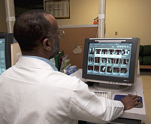
| (Photo credit: Wikipedia) |
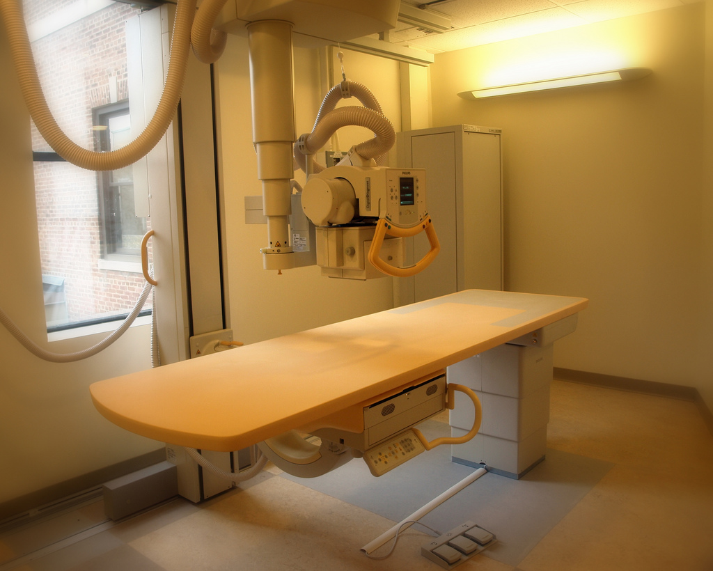
So I have defined the beginning of this journey. I have an endpoint in mind too: A dermatology specific IOD recommendation for WG-19 DICOM. Don’t worry if you don’t understand the endpoint now. Suffice to say that we will partly follow the specialty with a well defined imaging standard; Radiology, and modify their tools for our domain. At this juncture, the path to that endpoint is less clear to me too! Technical parts of this series will be on my informatics blog.
Here is the link to the wiki page, where I shall collect all the resources, and this is where we shall reach our destiny with your comments and contributions! I have given the name DICODerm (Digital Imaging and COmmunications in Dermatology) for this project. So if you tweet or discuss in #DsB (Dermatologists Sans Borders), please use the hashtag #DICODerm and feel free to follow me @beapen
Here is a useful preliminary solution to your image organization problems! Dermatology Image Tagger.
Disclaimer: I am not a member of NEMA, Medical Imaging and Technology Alliance or WG-19. The opinions mentioned here and internally hyperlinked pages are my own. External sites are hyperlinked in good faith, but does not mean endorsement.

- Machine learning-based BOTOX API - April 11, 2023
- Skinmesh: Machine learning for facial analysis - November 10, 2020
- Free Dermatology EMR for Machine Learning and Artificial Intelligence - January 2, 2020


Bell I am an avid fan of digital photography for my patients. It helps me many ways. Intact I have TB’s (terrabytes) of pics of my patients over last 10 years. I use nikon D90 with the various lenses. Pics help me many ways. .
1. For me to go back and compare the pre condition of skin
2. In case when patient claims they did not have the problem like hair prior to laser.
And so on and so on. ……
However I would say the whole essence of cosmetic practice is in actual listening and feeling. I love to chat with my patients. Listen to what bothers them. Most of the time it is nothing that cm be captured on camera. It’s just their mind. It’s too feel. That attention to a minute little microscopic speck on their skin makes them happy.
Well u could go on writing but I don’t want to divert the topic of discussion. Open to comments. …..
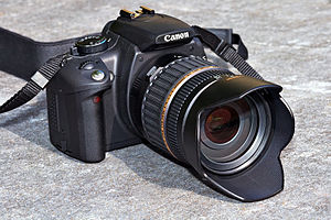
My Canon DSLR is my shadow, capturing my patients’ initial complaints & how I hv helped sorted them out.
It also helps me improvise my techniques from time to time. In fact, like you Apratim, I have now separated my music backup TB to a new one.
Thanks @Dr Apratim Goel & @Dr Geraldine Jain for the comments:
I will just give you a summary of what I am trying to do. Radiology uses a standard called DICOM (complicated yet effective) and have systems to store, retrieve and transfer images called PACS. Modifications of this system is being used in Pathology and Ophthalmology. Dermatologists (AAD) started working on a dermatology standard within DICOM, but never progressed. They pertinent question is: do we really need this standard? I need your help to sort things out.
I have few questions about your experience with clinical photographs:
1. Do you get confused about the patient’s identity and the clinical history when you review your old photographs? Do you have a filing system?
2. Do you have a system to know the time difference between two photographs?
3. Do you feel constrained because your storage device can only be in one of your clinics?
4. Do you rely on assistants to take photographs? Will it be helpful if you have a system that reminds them about the standard views to capture?
5. Do you share images with your colleagues? If you do, what all information do you send along with the image? Will it be helpful if those information is present in the image itself?
And finally, to the teledermatology society of India:
1. Do you have any successful implementations in India? What are the challenges you have faced?
I have all the above problems as well as many more. 1 to 4 are highly applicable. Not 5 for me.
Thanks Apratim. Please add your other problems too, when you have time.
Bell whenever I attend conferences I am never happy about the quality of pre post pics. Infact I personally take so much pains to put up the right pre post for comparison and am still not very happy with the quality of my own pics. But I see so many national as well as international speakers with horrible pre post. They are either distant angles or over exposed or near our far etc etc. ..I use the omnia system in both my clinics but that can only be used on face. So if something comes up for managing this problem it will be a boon.
Also the storage and identification of old pics esp of body parts is a huge issue. Now I have kept someone who only managed pics. But then half of the times she is running after the people who have taken the pics. As the therapists and doctors take pics during treatment in rooms. She keeps being them to identify pics.
We store our pics in tb external hard drives. So far 3 are full. But if you ask me to pull out 100 pre post of any treatment which would qualify for publication, I am sure I will start fumbling. You know what I mean.
I use a software system for patient data management and am very happy with it. It is Internet based. But can’t load pics on that. Also I am always concerned about the privacy of my patients as well.
Thanks a lot Apratim for this post. It summarizes all our challenges (Capture, Organizing, Storage, Retrieval and Transmission). If we think about radiologists, they face all these problems at a much larger scale. But they have very efficient systems for managing their resources. The idea is to adapt their tools for our needs (so that we don’t have to rediscover the wheel). I will need your help again when I start working on the technical details mainly to define the standards for cosmetic dermatology. I need help with the photography techniques too:
1. How do we standardize the views?
2. Can we standardize the parameters in a vendor neutral way?
Feroze, Ashique, Rashmi Suneil : your views please!
If we can come up with an internationally recognized standard, Nikons or Canons of tomorrow might have an option for dermatologists!! OK, that may be too far fetched 🙂
A lot of work is being done in this area…the pioneers seem to be fotofinder and canfield. One of the best things I have seen recently is the ‘ghost’ feature of fotofinder machines, where the screen shows the ‘ghost outline for the post-images….you just have to place the patient within the outline.
Thanks Feroze . The problem with vendor specific standards is interoperability. Vendors don’t share standards because they want everybody to buy their systems. Any encoding algorithms beyond JPEG?
True…would be nice to make some kind of open source software for the same…wouldn’t need that many hi-fi features..
Hmmm… My idea is to set the standards initially. Software will follow. In fact DICOM has already set the standard, with applications beyond radiology. We just need to tweak it to our needs. The tweaking needed is in fact very minimal (I feel). Then many of the existing DICOM viewers may support our needs with minimal alterations. But the question is: Are we ready for a change from ‘plug your card – save it in picasa’ to a more organised image management workflow? Do we need it? I may be biased!
Bell of course we are ready. I would rather say we are late.
I would be happy to be of any assistance Bell. The challenges apart from the ones discussed so far are. ..
1. If there is a cloud based system which does not occupy the disk space is better. Hard disks can also get corrupted. I have lost lot of data.
2. With multiple clinics I face a problem of viewing the pics at different clinic from where it was taken
3. Lots of patients want to see their pre and post pics as comparison. That’s My dreaded day. It takes so long to choose the worst pre and best post.
4. Some ask to give them a copy of their pics. That’s even worst. Neither can I say yes nor refuse. Yes will mean too much time and no would mean something else. Intact couple of times when we tried to delay this procedure in repeated requests of patients they said that why the hell do you take our pics on each visit if you can’t provide us the results on each visit. Some even refused for pics to be taken further
5…….. my list is endless Bell. And you can see how passionate I am about pics and how frustrated it leaves me.
we were actually using DICOM for our teledermatology consultation back when I was in Amrita, Kochi…and you’re right, the comfort level was much more for JPEG…
Thanks Apratim. Again that summarizes many of the problems that we can solve. In fact solutions for your problems exists in other specialities. We just need to adapt it to our needs:
http://en.wikipedia.org/wiki/Picture_archiving_and_communication_system
Feroze : The problem with DICOM now is that it is not fine tuned for dermatology. In technical terms we use Secondary Capture (SC) and Visible Light (VL) IODs for encoding dermatology pictures. Once we have a dermatology IOD, vendors may come out with devices supporting that. (Pathologists have already done that) DICOM is not just about encoding, It defines a whole lot of other things. But the icing on the cake is that we can use existing PACS systems for store-and-forward teledermatology!
I enjoyed reading your blog Dr Eapen. I’m quite impressed with the Antera 3D Imaging system though I’m still finding my way through it and have only recently started comparing before and after pics in terms of melanin content and scarring. I’ve been experimenting with the ‘diagnose with photograph’ idea but I find that unless I can touch, feel, squeeze or at times, look at a lesion in tangential lighting, I end up second guessing my own diagnosis. Online consultation and treatment does not work very well in dermatology (MY two cents).
Would like to thank Dr Apratim Goel as well, as her comments have been an education on their own. Standardization is my biggest nightmare, not just with Nikon but with computerized 3D imaging systems as well
Thnx Bell for this very invigorating discussion. I face all problems 1-4 & even more. Even tho’ I hv a studio & staff to take pics, I still end up doing them myself.Of course we need better “Image management’.
There are multiple companies abroad that now provide the tools for in-office photography for the practicing Dermatologist. They provide the equipment as well as the software to take & store patient photographs efficiently & accurately. The images are stored in individual patient files, in a safe & Health Insurance Portability & Accountability Act ( HIPAA) compliant manner.
A friend introduced me to Frankfort’s position( For standardization). If anyone interested, pls let me know.
Thnx for the gud discussion. Your endeavor is laudable, Bell Eapen
@Sunaina: I fully agree that diagnosing from digital images is different from diagnosing the same on a patient. I feel digital diagnosis will emerge as a ‘specialized skill’ that requires additional training, just like cosmetic dermatology carved out its niche. The right tools will enable this transition. BTW I took some time to figure out who this Dr Eapen is Bell is just fine!
@Geraldine: The problem with vendor specified standard is the issue of interoperability. I never knew about ‘Frankfort’s position’. Just now googled to find out. Would love to know more about it. Thanks for the support. Shall post more on the way ahead on the blog.
Breaking the monopoly of vendor specific systems & fine tuning a software or programme to suit our needs is what I think you are looking at , Bell. I remember how I would pick up tips from my family (which boasts of 2 prof Photographers ) & a friend whose shots are acclaimed world wide.
You stand to go down in the annals of the history of Dermatology for this breakthro’. I’m sure Apratim will agree with me.
Frankfort horizontal position helps to standardize facial photos. Here the infraorbital rim is at the same level as the supratragal notch. Full-face frontal, right & left 45 degree oblique,& right & left lateral photos are typically obtained in a facial series.
If any one is interested in tips for beginners, I’ll only be glad to share.
@ Geraldine: Thanks a lot for the kind words of encouragement. But the fact is, the melodrama in the blog is intentional, and what I am setting out to do is far less exciting! In brief, when DICOM standards were introduced, they knew its application in other specialties including dermatology. They have a workgroup for dermatology standards called WG-19, and they started working on a dermatology standard in 2008. But they did not progress much probably because there was very little public interest. I am just trying to see whether things have changed in the last 5 years and to wake them up by putting our 2 cents in. That 2 cents will be worth far more, if it comes from a strong group. Thanks for information on Frankfort position.
In fact I wanted to introduce this document with more melodrama But here it is, the summary of the problems explained by a true expert (from the minutes of standards committee meeting).: http://medical.nema.org/…/DICOM-Dermatology…
There’s a lot of change in the last few years & believe me, it’s need induced. So U hv a battalion backing you with their last dime. GO For It!!
DR Madden has 2 points for us to chew on
Whether diagnostic performance would improve if better imaging and analysis systems were available to the dermatologist? In addition, whether the improvement in diagnostic accuracy would lead to savings at least equal to the cost of the hardware.
Can we hv some views of our learned collegues?
I don’t know if I have ever had a diagnostic confusion in my practice Geraldine. However you will have to excuse me as I have mostly cosmetic practice with lasers and injectables. May be this will help in dermatology practice.
The way I look at this is.
1. It helps me in proper data storage and which is easy to access too. Since I run multiple clinics hope the data will be accessible from multiple sites.
2. Pre post easy to show to patients on follow up discussions
3. For publication and presentations it will be a great asset.
4. I do train doctors at my clinic on various procedures. This would be a great value add to them.
5. For my own learning.
However I must confess I don’t understand lot of technical language that Bell and Geraldine are speaking in this discussion. Am I completely on different track ?
@ Apratim: Geraldine is referring to the link I posted above to the minutes of a standard committee meeting. The practical points you mentioned are vital and will be the main advantage of such a system and the reason for its acceptance. In fact you are on the ‘right’ track than me here.
@ Geraldine: I am a firm believer of what he alludes to! The pixel patterns of a standardized picture may contain hitherto unidentified diagnostic information. Maybe in the future, standardized digital photography may become a diagnostic test like biopsy! If that happens, definitely it will be commercially viable.
@All: I am curious to know your views on another statement he has made: “The view of the individual clinician is very different from that of the enterprise.”
I too think there are multiple things being discussed- one issue most dermatologist face is the archiving/retrieving of images and the pre-post standardization. DICOM will definitely help a lot in the archiving and easy retrieval….with the advantage that it can be futher linked to electronic medical records both for in-house use and teledermatology The pre-post standardization is where the frankfort position, ghost image technology, photography frames etc come in. The third aspect is the standardization of colors in the image. That’s one thing I know Bell has already done some work on..and this is pretty essential for ‘digital diagnosis’…interesting bit of info from Brian Madden…keep us posted and let us know if we can help in anyway.
Are there any studies on automated diagnosis based on ‘pixel pattern’?
@Feroze: Nothing I am aware of at present. But I feel it may be possible with algorithms like ‘Neural Networks’. If you train them on enough standard pictures, maybe they can find patterns that cannot be explained, but still useful for prediction. If not a clinical diagnosis, at least a pathological one like inflammation/hyperkeratosis/pigment incontinence. I believe there are systems for pathology slides. What do you think?
Sorry for chipping in late, but Apratim U r on the right track. A system like DICOM can be useful only with valuable inputs from clinicians like you & the rest of us `who value Photo-documentation.
DICOM will add to bring Teledermatology to the forefront.
@ Feroze: (On the first point) In fact my idea is to include pre-post standardization also as part of DICOM dermatology standard. Technically ‘post’ will refer to its ‘pre’ for easy tracking. Standard views will be part of a single object, though some views may be null (but defined). Ultimately description of primary and secondary lesions will also be encoded along with the image. So DICODerm will be a much more comprehensive standard specific for dermatology and it will set the stage for dermatology specific image capture systems to communicate seamlessly with existing PACS systems.
Right…what I meant is that even the pre has to be in a standard format in terms of patient position, lighting etc.That would really need some kind of photographic frame like the visia/omnia/fotofinder ones. DICODerm (nice name by the way!) does seem a really exciting prospect!
@Geraldine: Being HIPAA compliant is really complicated. The best way to make the capturing device compliant is to avoid secondary storage in the device. If the storage system follows standards, it will be much easier to make it HIPAA compliant. See this link: http://www.ncbi.nlm.nih.gov/pmc/articles/PMC3045193/
The discussion, Geraldine has posted above/below takes me to last point I wanted to discuss: The importance of encoding clinical history and preliminary/pathological diagnosis. DICOM already has standards for capturing a radiologist’s report and associating it with the image (DICOM – SR). We can actually tweak it for encoding history and diagnosis.
@Feroze: Right. It will be great if you can work on standardizing capture. Maybe they already have guidelines to define capture. Because positioning, intensity etc are important in X-ray, CT and other radiology image capturing devices. Maybe X-ray standards will be more in line with our needs as positioning is manual. Full DICOM text >3000 pages is available here http://medical.nema.org/standard.html
Right. Thnx
Will do and get back…..only 3000 pages?:)
I think part 3 (Information Object Definitions) will be the most relevant.
Hi All: I have started to update details on the wiki page http://gulfdoctor.net/wiki/index.php?title=DICOD&lang=en (This is a wiki, you can register and edit this page on your own, Remember to add your name). Feroze, Ashique, Please advice on Positioning Attributes and Acquisition Attributes. Apratim, Geraldine & Sunaina, What all data you want embedded in an image? in other words, what are your searches generally based on? (Name, diagnosis etc.) It will be great if we can get more support for this.
Reopening #DICODerm @ Feroze Do you think we can formulate a guideline for image acquisition: aperture, shutter speed, ISO etc??
Geraldine / Apratim / Sunaina : How do you search for an image from your collection?
Search based on patient’s name or ID
Search based on patient’s date of visit
Search based on diagnosis (eg: All images of lichen planus)
Search based on lesion (eg: All vesiculobullous lesion photos)
What else is practically important?
Bell I used to earlier do it according to the indication or treatment done. But then it became cumbersome as a patient would do multiple treatments and it would be difficult. Do now we make a folder by his or her name and irrespective of the treatments done, we put all pics there. But in that we make sub folders date wise.
Retrieving images based on patients’ name, date of visit or id is like looking for a needle in a haystack in my multiple setups.
A primary folder is made based on diagnosis & a number allotted (eg 1 for acne ), sub-folders follow by the patient’s name & all images go there. Now, I have access to the images in any of the branch the patient chooses to visit.Date of visit is automatically stored in all images but does not contribute to the retrieval.
We also make sure to have back up of backup of backup & one backup is stored away from all 3 centers, at a friend’s place.The 3rd back up is updated every month.
A separate 1TB hard drive is with me all the time where I store cropped images for my presentation purposes.
We create different folders based on procedure done eg hair removal laser, co2 laser etc, also based on indications say pigmentation, nevi, complications etc. Each pt has their own folder within that folder. We also label name, date etc before taking pic. Data storage and retrieval is not an issue, standardization is
Certain patient filing softwares also have provision to save images directly to pt file, like the updated medtrix system
@All. Thanks a lot. Geraldine’s backup routine is amazing! Let me reformulate the question in another way.
Imagine, you have received an image from a colleague through email. (There is only image, nothing else). What other information do you seek from your colleague, to file that image in your system?
OR, Imagine, you have a google like search system for retrieving your images. What are the possible search options you would use? Other than name, id, diagnosis, treatment.
If we achieve our goal: Your camera/device will tell your assistant how to capture the image, Your device will know, where and how to send it, Your filing system will know how to file it so that you can search and find that image later. Your image viewer will know how to display it accurately. If you email it to a colleague, his/her filing system will know how to file it! Your EMR (and your colleague’s EMR) will know the source of the image! (Even if all of us are using different cameras, filing software, EMRs and assistants ) That is what happens in radiology now.
sorry for joining in late. The problem is that the aperture/shutter speed /ISO will depend a lot on other variables also – especially, but not limited to lighting and the type of camera/lens used. How can we factor this in?
hmmm. @ Feroze Please see this document when you have time. This is an Ophthalmology IOD. I think it will give us a framework.
http://one.aao.org/asset.axd?id=5a1d1f07-5381-4404-83d9-e51285784781&t=634962790626070000
interesting read…would be great if we could have something like that mentioned on page 18…a lot of this would be covered in the EXIF data of the photograph (Though i realize that as mentioned in page 32, the devices and illumination in opthal are a bit of a different ball game compared to dermato photography ). Does dicom directly link to the EXIF data of the image? If so for a standard lighting and distance the rest of the settings – iso, aperture, shutter speed etc. will automatically be obtained. Not sure if I am making sense to even myself:)
DICOM file format is glorified EXIF, So we can write a wrapper that extracts Standard EXIF data to DICOM. ( http://www.sno.phy.queensu.ca/~phil/exiftool/ ) But future dermatology specific devices could directly output DICOM. We shall use the ophthal IOD as a template and modify the wiki page accordingly.
Here is a useful preliminary solution: Dermatology Image Tagger available from http://gulfdoctor.net/dit/
Excellent goods from you, man. I’ve understand your stuff
previous to and you’re just extremely fantastic.
I really like what you have acquired here, really like what you are stating and
the way in which you say it. You make it enjoyable and you still take care
of to keep it wise. I can’t wait to read
far more from you. This is really a terrific site.