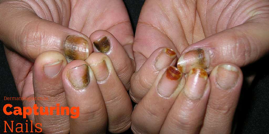
The above image shows finger nails taken in direct view and the limitation is evident!
Documentation of nail disorders by clinical photography has certain practical difficulties. In the toes nails, it is very easy as all the nails are in the same plane when the feet is placed on a flat surface. For the fingernails one may be able to do it by asking the patient to hold the fingers in various positions, like clenching, folding etc. [Figure shown at the beginning of this page]. However the procedure has its own limitations. The main drawback is that the corresponding images cannot be standardized. The very same patient may not be able to hold the fingers in the same order next time he comes for review. It is especially important when large/multicentric studies are undertaken to evaluate nail disorders requiring frequent imaging of nails in serial succession. We have found a simple technique that can overcome this problem and this could standardize the concept of imaging in fingernail disorders. This has been published as a pearl In the Journal of American Academy of Dermatology [JAAD] 1.
[Please get in touch with authors if you don’t have access to the pdf]- Imaging in nail disorders: An easy way out. - December 25, 2014
- Imaging in nail disorders: An easy way out. - December 25, 2014
References:
- Ashique, Karalikkattil T. et al. Clinical photography of nail diseases: A simple method to include all fingernails in a frame. Journal of the American Academy of Dermatology , Volume 72 , Issue 1 , e25 ↩

Leave a Reply