
In a small town called payyanur in Kerala, INDIA, three doctors were suspended for allegedly sharing a labour room video of triplets being delivered by C-section on social media. The powerful local media rebuked the medical community for their irresponsible behaviour.
Is taking clinical photos on your mobile second nature for you? Do you share them on Facebook?
Dermatology is a speciality that depends on visual cues. Photography can even be considered therapeutic in dermatology, since it is an important part of disease monitoring. Social media like facebook is an important and popular platform for crowdsourcing diagnosis. This is especially important for those practicing in resource deprived areas. How can this be achieved without compromising patient privacy?
#Facebook post, removed records among latest #patient #privacy #breaches http://t.co/ZKgIhaf5 #HCSM #Healthcare
— Brad Justus (@Brad_Justus) January 4, 2012
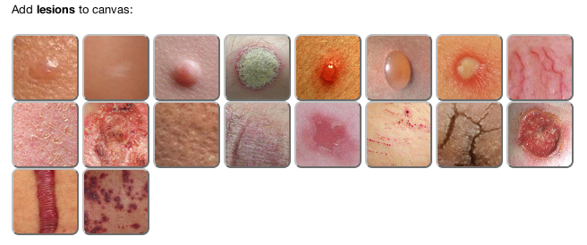
I have developed a tool for visual diagnostic information encoding. The tool called LesionMapper(TM) captures information such as location, distribution, progression and severity using pre-defined representative lesion images and icons. It also provides the capability to create lesion icons from existing clinical images. Without further ado here is the link to LesionMapper(TM): http://skinhelpdesk.com/lesionmap.html
 |
| Lesion Canvas for LesionMapper(TM) |
The instructions tab has details on how to use it. Below I have added basic steps to add a lesion from your photograph. Please note that using LesionMapper(TM) does not guarantee anonymity. It is your responsibility to ensure that you have obtained the necessary consent. If the preset images and icons are insufficient, you can crop the lesion from the clinical image itself as below.
- Go to http://skinhelpdesk.com/lesionmap.html
- Click on Upload Image 1 (Blue Button)
- Upload your image. Select only the lesion and follow instructions. (In the new window)
- Click on Upload 1 (White button). Image will be added to canvas. If not press the button again.
- Drag the image to the area of involvement. You can resize the image if needed.
- Click on the Download button (A small button right below the image). You can also save it on the site. You will be provided with an ID that can be used to render the image by clicking on Load.
Please share below to see the reference to medico-legal considerations in dermatology photography.
[sociallocker id=”771″]
Reference:
Kunde, L., McMeniman, E. and Parker, M. (2013), Clinical photography in dermatology: Ethical and medico-legal considerations in the age of digital and smartphone technology. Australasian Journal of Dermatology, 54: 192–197. doi: 10.1111/ajd.12063 (http://www.ncbi.nlm.nih.gov/pubmed/23713892)
[/sociallocker]
- Machine learning-based BOTOX API - April 11, 2023
- Skinmesh: Machine learning for facial analysis - November 10, 2020
- Free Dermatology EMR for Machine Learning and Artificial Intelligence - January 2, 2020
Leave a Reply